Beranda
/ Foot Muscles Mri : Anatomy Of The Foot And Ankle Mri : Anatomical structures of the ankle and foot and specific regions (major joints) are visible as dynamic labeled images.
Foot Muscles Mri : Anatomy Of The Foot And Ankle Mri : Anatomical structures of the ankle and foot and specific regions (major joints) are visible as dynamic labeled images.
Insurance Gas/Electricity Loans Mortgage Attorney Lawyer Donate Conference Call Degree Credit Treatment Software Classes Recovery Trading Rehab Hosting Transfer Cord Blood Claim compensation mesothelioma mesothelioma attorney Houston car accident lawyer moreno valley can you sue a doctor for wrong diagnosis doctorate in security top online doctoral programs in business educational leadership doctoral programs online car accident doctor atlanta car accident doctor atlanta accident attorney rancho Cucamonga truck accident attorney san Antonio ONLINE BUSINESS DEGREE PROGRAMS ACCREDITED online accredited psychology degree masters degree in human resources online public administration masters degree online bitcoin merchant account bitcoin merchant services compare car insurance auto insurance troy mi seo explanation digital marketing degree floridaseo company fitness showrooms stamfordct how to work more efficiently seowordpress tips meaning of seo what is an seo what does an seo do what seo stands for best seotips google seo advice seo steps, The secure cloud-based platform for smart service delivery. Safelink is used by legal, professional and financial services to protect sensitive information, accelerate business processes and increase productivity. Use Safelink to collaborate securely with clients, colleagues and external parties. Safelink has a menu of workspace types with advanced features for dispute resolution, running deals and customised client portal creation. All data is encrypted (at rest and in transit and you retain your own encryption keys. Our titan security framework ensures your data is secure and you even have the option to choose your own data location from Channel Islands, London (UK), Dublin (EU), Australia.
Foot Muscles Mri : Anatomy Of The Foot And Ankle Mri : Anatomical structures of the ankle and foot and specific regions (major joints) are visible as dynamic labeled images.. In addition, an image of all the muscles of the back and plantar part of the foot, all tendons and tendon ligaments, blood vessels and nerves are obtained. The most common ossicle is the os trigonum, which is a prominent unfused apophysis of the lateral tubercle of the talus. Adductor hallucis is anatomically located in the central compartment of foot, but the muscle is functionally grouped with the medial plantar muscles of foot because it acts on the great toe (hallux). Those fibers of the most medial and largest belly are… Routine ankle magnetic resonance imaging (mri) tests involve taking images of the foot and ankle in the axial, coronal, and sagittal planes parallel to the tabletop(2).
Denervation changes in muscles early. One of the large muscles of the leg, it connects to the heel. Magnetic resonance imaging, otherwise known as mri, uses a combination of magnetic fields and radio waves to take images of the internal structures of your body. Muscle damage may cause muscle pain and muscle weakness may cause difficulty lifting the arms above the shoulders, climbing stairs, or arising from a sitting position. Related posts of foot muscle anatomy mri muscle anatomy trivia.
Foot Radiological Anatomy Shorouk Zaki from image.slidesharecdn.com Trauma effects of direct injury or tear denervation injury: Magnetic resonance imaging, otherwise known as mri, uses a combination of magnetic fields and radio waves to take images of the internal structures of your body. Denervation changes in muscles early. Learn about foot and ankle mri. Accessory muscles are isointense to skeletal muscle on all pulse sequences, and can insert by fleshy muscular or tendinous insertions. Plantar plate of the foot: Findings on conventional arthrography and mr imaging. The adductor hallucis has two heads:
Learn about foot and ankle mri.
The most common ossicle is the os trigonum, which is a prominent unfused apophysis of the lateral tubercle of the talus. One of the large muscles of the leg, it connects to the heel. Findings on conventional arthrography and mr imaging. The three plantar interossei muscles adduct the 3 rd, 4 th and 5 th toes toward the long axis through the 2 nd toe. Learn about foot and ankle mri. Routine ankle magnetic resonance imaging (mri) tests involve taking images of the foot and ankle in the axial, coronal, and sagittal planes parallel to the tabletop(2). A mri of a foot can show detailed images of bones, cartilage, tendons, muscles, blood vessels, and ligaments, which allows your healthcare provider to identify where the source of the pain is located and what further damage it has caused or is causing. An extremity mri is a type of scan used specifically for diagnostic imaging of the arm, leg, hand, or foot. In addition, an image of all the muscles of the back and plantar part of the foot, all tendons and tendon ligaments, blood vessels and nerves are obtained. Mri findings of acute turf toe: Mri of the ankle and feet Denervation changes in muscles early. In the foot and ankle many accessory ossicles can be seen.
Plantar interossei (foot) dr yuranga weerakkody ◉ and dr geon oh et al. The aim of this review is to provide the reader with a comprehensive overview of the magnetic resonance imaging (mri) characteristics of the most common benign and malignant soft tissue neoplasms which occur around the foot and ankle. This imaging technique assesses the ligaments and tendons, neurovascular structures (tarsal tunnel and plantar fascia), and the osseous structures(19). Those fibers of the most medial and largest belly are… Muscles of the foot muscle origin insertion nerve supply extensor digitorum brevis distal part of the lateral and superior surfaces of the calcaneus and the apex of the inferior extensor retinaculum as the fiber bundles extend distally, they become grouped into four bellies.
Advanced Mr Imaging Techniques For Differentiation Of Neuropathic Arthropathy And Osteomyelitis In The Diabetic Foot Radiographics from pubs.rsna.org Mri is an ideal method for identifying areas of muscle atrophy and fatty infiltration. The peroneus quartus muscle is more common, presenting in 13% to 22% of the population. Mri of the ankle and feet The studies were performed on a variety of magnets ranging from 0.2 to 1.5 t between march 15 and july 22, 2006. Muscles that move the foot and toes. The aim of this review is to provide the reader with a comprehensive overview of the magnetic resonance imaging (mri) characteristics of the most common benign and malignant soft tissue neoplasms which occur around the foot and ankle. The adductor hallucis has two heads: This imaging technique assesses the ligaments and tendons, neurovascular structures (tarsal tunnel and plantar fascia), and the osseous structures(19).
Ultrasonography (us) affords high spatial resolution of muscle but is less sensitive than magnetic resonance (mr) imaging for mild edema and early myopathy.
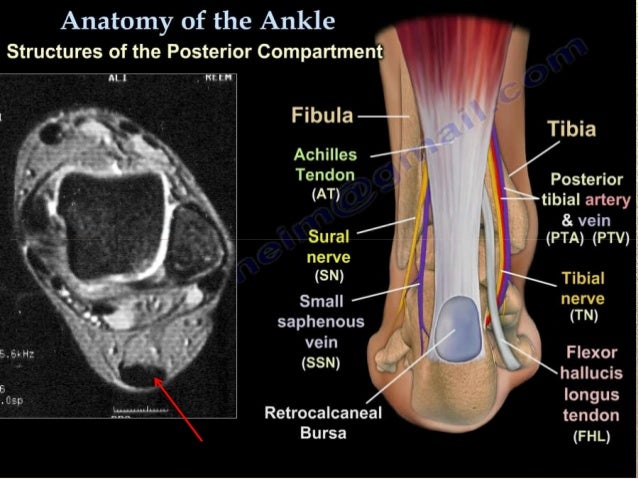
Anatomical structures of the ankle and foot and specific regions (major joints) are visible as dynamic labeled images. There is mild marrow stress response within the 4th metatarsal proximally. The intrinsic muscles of the foot are key contributors to foot function and are important to evaluate in lower limb disorders. Coronal images are perpendicular to the long axis of the metatarsals. Learn about foot and ankle mri. Mri of the ankle and feet Denervation changes in muscles early. 9 yao l, do hm, cracchiolo a, et al. Muscles that move the foot and toes. Findings on conventional arthrography and mr imaging. Both muscles are innervated by the deep fibular nerve. Magnetic resonance imaging, otherwise known as mri, uses a combination of magnetic fields and radio waves to take images of the internal structures of your body. With a muscle injury, for example, mri images often show a bright signal indicating that there is more water in the muscle, which is a sign of injury.
This small, thin muscle is absent in about. 23,25 mri at the level of the malleolus demonstrates the muscle as. Coronal images are perpendicular to the long axis of the metatarsals. Muscle damage may cause muscle pain and muscle weakness may cause difficulty lifting the arms above the shoulders, climbing stairs, or arising from a sitting position. Both muscles are innervated by the deep fibular nerve.
25 Magnetic Resonance Imaging Of Foot And Ankle Pathology Musculoskeletal Key from i2.wp.com The three plantar interossei muscles adduct the 3 rd, 4 th and 5 th toes toward the long axis through the 2 nd toe. 23 it can originate as a separate muscle from the fibula or from the peroneus brevis or longus muscles and inserts onto the peroneal tubercle or retrotrochlear eminence of the calcaneus. The aim of this review is to provide the reader with a comprehensive overview of the magnetic resonance imaging (mri) characteristics of the most common benign and malignant soft tissue neoplasms which occur around the foot and ankle. An extremity mri is a type of scan used specifically for diagnostic imaging of the arm, leg, hand, or foot. The peroneus quartus muscle is more common, presenting in 13% to 22% of the population. Muscles that move the foot and toes. It flexes and extends the foot, ankle, and knee. Routine ankle magnetic resonance imaging (mri) tests involve taking images of the foot and ankle in the axial, coronal, and sagittal planes parallel to the tabletop(2).
Accessory muscles are isointense to skeletal muscle on all pulse sequences, and can insert by fleshy muscular or tendinous insertions.
Findings on conventional arthrography and mr imaging. The muscles of the dorsum of the foot are a group of two muscles, which together represent the dorsal foot musculature. Muscle damage may cause muscle pain and muscle weakness may cause difficulty lifting the arms above the shoulders, climbing stairs, or arising from a sitting position. In the foot and ankle many accessory ossicles can be seen. 23 it can originate as a separate muscle from the fibula or from the peroneus brevis or longus muscles and inserts onto the peroneal tubercle or retrotrochlear eminence of the calcaneus. The machine uses radio waves and a magnetic field to generate images of the inside of the extremity in order to diagnose problems with the muscles, bones, joints, nerves, or blood vessels. Trauma effects of direct injury or tear denervation injury: Mri is the choice of modality for further imaging the ankle and foot after obtaining initial radiographs. Mri findings of acute turf toe: One of the large muscles of the leg, it connects to the heel. Magnetic resonance imaging, otherwise known as mri, uses a combination of magnetic fields and radio waves to take images of the internal structures of your body. Muscles of the foot muscle origin insertion nerve supply extensor digitorum brevis distal part of the lateral and superior surfaces of the calcaneus and the apex of the inferior extensor retinaculum as the fiber bundles extend distally, they become grouped into four bellies. Learn about foot and ankle mri.